fbpx

Contact Number

+91 9584 889068

Canon Fundus Camera

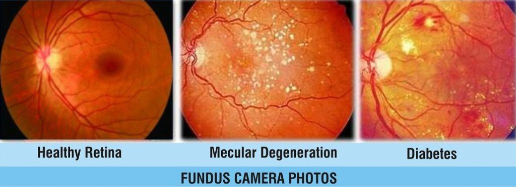
This highly specialized 35mm or digital camera has special, high-powered lenses designed to focus on the structures of the back of the eye. It is basically an eye “microscope” with a camera mounted on it. Canon fundus cameraFundus photography is used to document and sometimes diagnose certain eye conditions. The word “fundus” describes the inside or back of the eyeball. A typical fundus photo would contain an image of the center of the very back inner wall of the eye – the retina. The optic nerve, macula and main retinal blood vessels are common structures seen in a typical fundus photo. Fundus photography is very useful to document the natural state of the back of the eye in order to give the retinal specialist a future reference to compare with during follow-up visits.

It is important to document the findings of most retinal diseases and conditions, especially diabetic eye disease findings, macular degeneration, epi-retinal membranes, macular holes and retinal tears and detachment.

The patient’s pupil must be well dilated in order to perform fundus photography. Dilating the pupils will cause blurry vision, especially reading vision, for several hours, if not, days – depending on the type and strength of dilating drop used. A diagnostic study called fluorescein angiography (FA) uses fundus photography to capture images of injected dye circulating throughout the retinal blood vessels. This is a very helpful study that can show leaking, swelling or circulation problems caused by various eye diseases like diabetic retinopathy and wet macular degeneration.

Book An Appointment

  • –Select Department–
  • Ophthalmology
  • Pediatrics
  • General & Laparoscopic Surgery
  • Obstetrics & Gynecology
  • Orthopaedics
  • Assisted Fertility
  • – Appointment Time –
  • 10:00am-2:00pm
  • 5:00pm-7:00pm

Digital Retina Cameras or Digital Fundus Cameras are used to capture images of the interior surface of the eye. Many of these Digital Retina Cameras boast features like angle variations, color, red-free and angiography imaging, high grade LCD monitors with easy to use features as well as DICOM compatibility and EMR interfacing.

These images of the retina, optic disc, macula and posterior pole are digital, which enables quick transfer and detailed image study as well as side-by-side image comparison and longitudinal tracking over time. These images are key in the identification and care of various eye diseases. Choosing the correct digital retina camera for a practices needs should be carefully considered.

Need Help? Chat with us